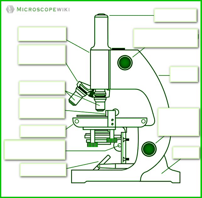
41 labeled diagram of microscope
A Study of the Microscope and its Functions With a Labeled Diagram ... These labeled microscope diagrams and the functions of its various parts, attempt to simplify the microscope for you. However, as the saying goes, 'practice makes perfect', here is a blank compound microscope diagram and blank electron microscope diagram to label. Anatomy of a Microscope | Microscopy Primer | Olympus LS The microscope illustrated in Figure 5 below was manufactured by Hugh Powell and Peter Lealand around 1850. The tripod base provided a sturdy support for the microscope, which many people consider the most advanced of its period. Parts of a Powell and Leland Microscope Diagram
PDF Electron Microscope Diagram Labeled April 28th, 2018 - Electron Microscope Diagram Labeled Electron microscope wikipedia an electron microscope is a microscope that uses a beam of accelerated electrons as a source of illumination as the wavelength of an Transmission electron microscopy an overview

Labeled diagram of microscope
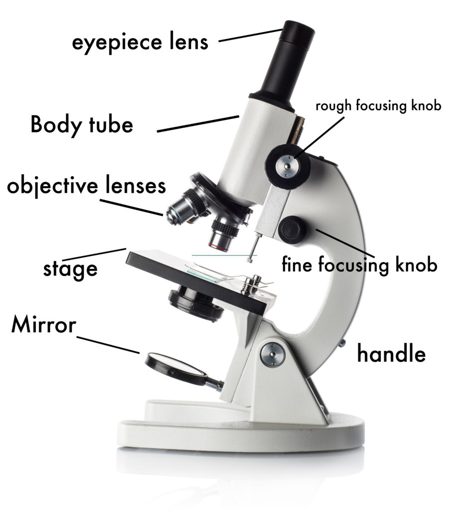
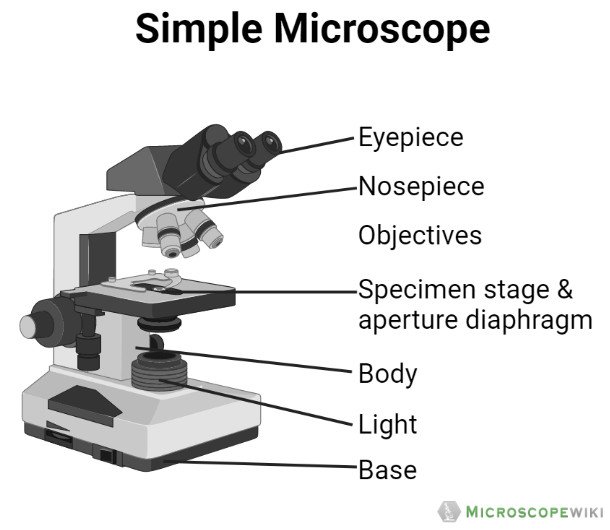
Parts of the Microscope (Labeled Diagrams) Simple microscope labelled diagram Image created with Biorender Tube/Body Tube It serves as the connector between the eyepiece/ocular and objective lenses. Objective lenses The lenses have varying magnifying power, which typically consists of 10x, 40x, and 100x. Simple Microscope - Diagram (Parts labelled), Principle, Formula and Uses A simple microscope consists of Optical parts Mechanical parts Labeled Diagram of simple microscope parts Optical parts The optical parts of a simple microscope include Lens Mirror Eyepiece Lens A simple microscope uses biconvex lens to magnify the image of a specimen under focus. Compound Microscope Parts - Labeled Diagram and their Functions Labeled diagram of a compound microscope Major structural parts of a compound microscope There are three major structural parts of a compound microscope. The head includes the upper part of the microscope, which houses the most critical optical components, and the eyepiece tube of the microscope.
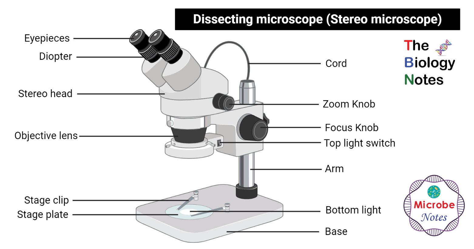
Labeled diagram of microscope. Types of Microscopes: Definition, Working Principle, Diagram ... These five types of microscopes are: Simple microscope Compound microscope Electron microscope Stereomicroscope Scanning probe microscope Simple Microscope A simple microscope is defined as the type of microscope that uses a single lens for the magnification of the sample. A simple microscope is a convex lens with a small focal length. Microscope Parts and Functions Microscope Parts and Functions With Labeled Diagram and Functions How does a Compound Microscope Work? Before exploring microscope parts and functions, you should probably understand that the compound light microscope is more complicated than just a microscope with more than one lens. Parts of Stereo Microscope (Dissecting microscope) - labeled diagram ... Labeled part diagram of a stereo microscope Major structural parts of a stereo microscope. There are three major structural parts of a stereo microscope. The viewing Head includes the upper part of the microscope, which houses the most critical optical components, including the eyepiece, objective lens, and light source of the microscope. Labeling the Parts of the Microscope | Microscope World Resources This activity has been designed for use in homes and schools. Each microscope layout (both blank and the version with answers) are available as PDF downloads. You can view a more in-depth review of each part of the microscope here. Download the Label the Parts of the Microscope PDF printable version here.
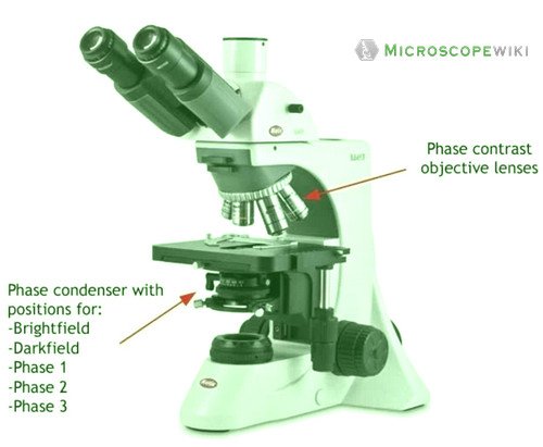
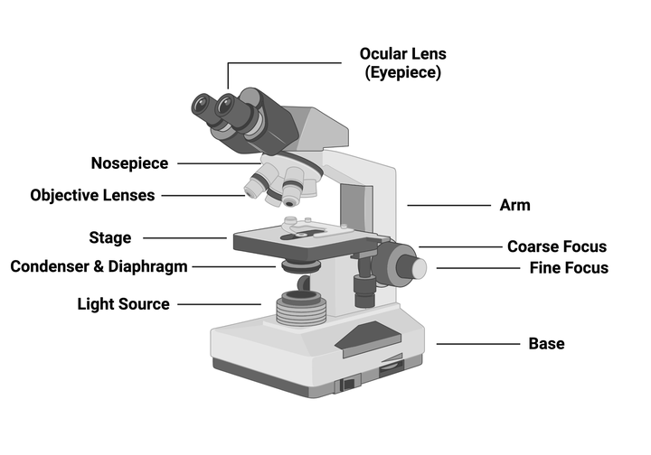
Microscope Types (with labeled diagrams) and Functions Phase-contrast microscope labeled diagram Phase-contrast microscope functions: Its applications areas include In cases where the specimen is colorless and is very tiny In biology to conduct cellular level examination of microorganisms that can't be visualized using the bright field microscopy Interference Microscope Light microscopes - Cell structure - Edexcel - BBC Bitesize The magnification of a lens is shown by a multiplication sign followed by the amount the lens magnifies. So a lens magnifying ten times would be ×10. The total magnification of a microscope is:... 16 Essential Microscope Parts: Names, Functions & Labeled Diagram Microscope Parts Labeled Diagram The principle of the Microscope gives you an exact reason to use it. It works on the 3 principles. Magnification Resolving Power Numerical Aperture. Parts of Microscope Head Base Arm Eyepiece Lens Eyepiece Tube Objective Lenses Nose Piece Adjustment Knobs Stage Aperture Microscopic Illuminator Condenser Lens Parts of a microscope with labeled diagram and functions There are three major structural parts of a microscope. The head comprises the top portion of the microscope, which contains the most important optical components, and the eyepiece tube. The base serves as the microscope's support and holds the illuminator. The arm is the component of the microscope that connects the eyepiece tube to the base ...
Parts of a microscope with functions and labeled diagram - Microbe Notes Figure: Diagram of parts of a microscope There are three structural parts of the microscope i.e. head, base, and arm. Head - This is also known as the body. It carries the optical parts in the upper part of the microscope. Base - It acts as microscopes support. It also carries microscopic illuminators. Compound Microscope Parts, Functions, and Labeled Diagram Compound Microscope Definitions for Labels Eyepiece (ocular lens) with or without Pointer: The part that is looked through at the top of the compound microscope. Eyepieces typically have a magnification between 5x & 30x. Monocular or Binocular Head: Structural support that holds & connects the eyepieces to the objective lenses. Dog Knee Anatomy with Labeled Diagram - AnatomyLearner Dog knee anatomy diagram. I hope all the diagrams of dog knee anatomy were helpful for you to understand it easily. If you need more diagrams on the dog knee joint, please follow anatomy learner on social media. You will get a notification if I update or publish any new diagrams on the dog knee in future. Frequently asked questions on a dogs knee Microscopy: Intro to microscopes & how they work (article) - Khan Academy In most cases, the part of a cell or tissue that we want to look at isn't naturally fluorescent, and instead must be labeled with a fluorescent dye or tag before it goes on the microscope. The leaf picture at the start of the article was taken using a specialized kind of fluorescence microscopy called confocal microscopy.
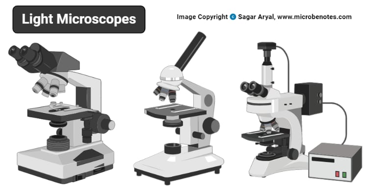
Light Microscope- Definition, Principle, Types, Parts ...
Microscope: Parts Of A Microscope With Functions And Labeled Diagram. List down the 18 parts of a Microscope. Ocular Lens (Eye Piece) Diopter Adjustment Head Nose Piece Objective Lens Arm (Carrying Handle) Mechanical Stage Stage Clip Aperture Diaphragm Condenser Coarse Adjustment Fine Adjustment Illuminator (Light Source) Stage Controls Base Brightness Adjustment Light Switch
How to Use a Microscope
Label the microscope — Science Learning Hub Use this interactive to identify and label the main parts of a microscope. Drag and drop the text labels onto the microscope diagram. diaphragm or iris base eye piece lens fine focus adjustment light source stage coarse focus adjustment high-power objective Download Exercise
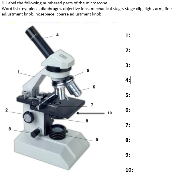
Solved 1. Label the following numbered parts of the | Chegg.com
Light Microscope- Definition, Principle, Types, Parts, Labeled Diagram ... Figure: Labeled Diagram of a Light Microscope. Types of light microscopes (optical microscope) With the evolved field of Microbiology, the microscopes. used to view specimens are both simple and compound light microscopes, all using lenses. The difference is simple light microscopes use a single lens for magnification while compound lenses use ...
Parts of a microscope with functions and labeled diagram
Microscope | Types, Parts, History, Diagram, & Facts The most familiar type of microscope is the optical, or light, microscope, in which glass lenses are used to form the image. Optical microscopes can be simple, consisting of a single lens, or compound, consisting of several optical components in line. The hand magnifying glass can magnify about 3 to 20×. Single-lensed simple microscopes can ...

Compound Microscope Parts – Labeled Diagram and their ...
Compound Microscope Parts - Labeled Diagram and their Functions Labeled diagram of a compound microscope Major structural parts of a compound microscope There are three major structural parts of a compound microscope. The head includes the upper part of the microscope, which houses the most critical optical components, and the eyepiece tube of the microscope.

Simple Microscope - Diagram (Parts labelled), Principle ...
Simple Microscope - Diagram (Parts labelled), Principle, Formula and Uses A simple microscope consists of Optical parts Mechanical parts Labeled Diagram of simple microscope parts Optical parts The optical parts of a simple microscope include Lens Mirror Eyepiece Lens A simple microscope uses biconvex lens to magnify the image of a specimen under focus.

Label the diagram of the microscope and explain the role of ...
Parts of the Microscope (Labeled Diagrams) Simple microscope labelled diagram Image created with Biorender Tube/Body Tube It serves as the connector between the eyepiece/ocular and objective lenses. Objective lenses The lenses have varying magnifying power, which typically consists of 10x, 40x, and 100x.

Microscope parts
Label microscope - Teaching resources

Compound microscope - their parts and function - Microscopy4kids

Microscope With Labels clip art | Microscope parts, Science ...
Solved Nikon Parts of the compound microscope Write the ...

Compound Microscope- Definition, Labeled Diagram, Principle ...

Parts of the Microscope with Labeling (also Free Printouts ...
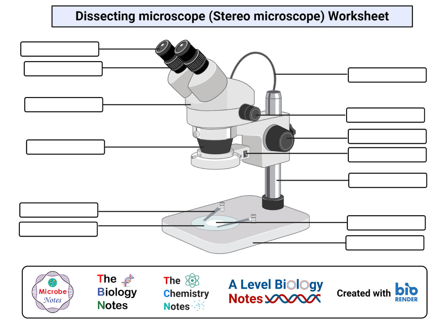
Dissecting microscope (Stereo or stereoscopic microscope ...
Figure 1.15 A labelled diagram of a light microscope | Boost
1.2: Microscopes - Biology LibreTexts
Parts of the Microscope (Labeled Diagrams) - Simple and ...
Parts of a Microscope Labeling Worksheet for Google Slides ...

Label the microscope — Science Learning Hub

Microscope Labeling
1.5: Microscopy - Biology LibreTexts

Labeled Parts Of A Microscope - ClipArt Best

Label The Microscope Parts! Diagram | Quizlet
What Is Light Microscope? – Microbiology Note
Simple Microscope - Diagram (Parts labelled), Principle ...

Parts of a microscope with functions and labeled diagram

Types, Parts and Functions of a Microscope

Compound Microscope Parts, Function, & Diagram | What is a ...
Microscope Types (with labeled diagrams) and Functions

Parts of a Microscope Diagram | Quizlet

Compound Microscope Principle, Parts, Diagram Definition ...
Light Microscope- Definition, Principle, Types, Parts ...

Label the Microscope Diagram | Download Scientific Diagram
Addgene: Using a Light Microscope Protocol

Labeling the Parts of a Microscope: Activity & Lesson Plan ...
Simple Microscope - Parts, Functions, Diagram and Labelling ...

Compound Microscope: Parts of Compound Microscope
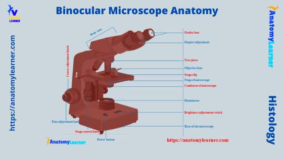
Binocular Microscope Anatomy - Parts and Functions with a ...
Label microscope - Teaching resources

Microscope Diagram - Label Diagram | Quizlet
Post a Comment for "41 labeled diagram of microscope"